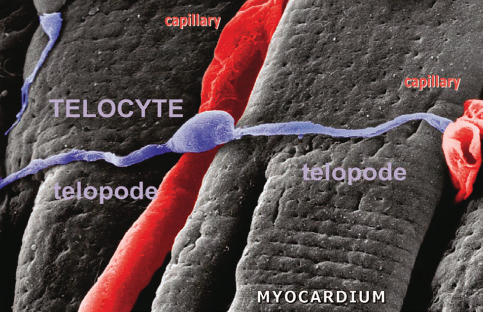
Representative scanning electron micrograph.Monkey left ventricular myocardium. The image shows a typical telocyte located across the cardiomyocytes. Another (possible) telocyte appears located near the cardiomyocytes (upper left). The 3D view reveals close interconnections of TCs with cardiomyocytes and capillaries. Note the cardiomyocyte striations and the openings of T tubules.
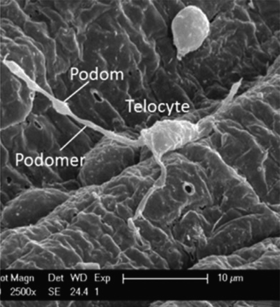
Scanning electron microscope image of a medium-sized artery in pig. A TC with three Tps and typical podomer and podom.
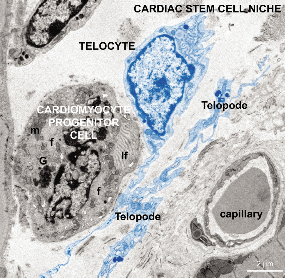
Electron microscopy images from epicardial stem cell niche showing more differentiated cardiomyocyte progenitors (CMP) with characteristic leptofibrils.
TC chaperone a low differentiated CMP with distinctive leptofibrils(lf), unorganized myofibrils (f), Golgi apparatus, and clusters of mitochondria (m).
Sanda M. Cretoiu, Laurentiu M. Popescu. Telocytes revisited. BioMol Concepts 2014
Yuli Kang a, Zaihua Zhu, Yonghua Zheng c, Weiguo Wan b, Catalin G. Manole, Qiangqiang Zhang. Skin telocytes versus fifibroblasts: two distinct dermal cell populations. J. Cell. Mol. Med. Vol 19, No 11, 2015